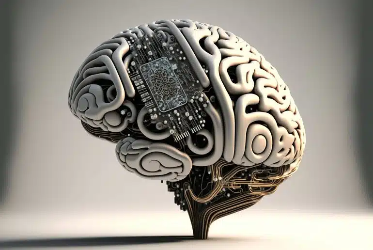
Researchers investigate brain region synchronization in order to assist control of brain-machine interfaces.
Just a few decades ago, the possibility of connecting the brain with a computer to convert neural signals into concrete actions would have seemed like something from science fiction.
But in recent years, some scientific advances have been made in this regard, through so-called BCIs (Bran-Computer Interfaces) that establish communication bridges between the human brain and computers.
A recent study by UPF continues to advance in this direction and makes new contributions to pursue this desired neuroscientific milestone.
The results of the study by the UPF Center for Brain and Cognition (CBC) are the subject of an article published on February 7 in the journal eNeuro, titled “Long-range alpha-synchronisation as control signal for BCI: A feasibility study,” jointly written by Martín Esparza-Iaizzo (UPF and University College of London), Salvador Soto-Faraco (UPF and ICREA), Irene Vigué-Guix (UPF), Mireia Torralba Cuello (UPF), and Manuela Ruzzoli (Basque Center on Cognition Brain and Language).
One of the main current challenges in neuroscience is the identification of brain signals which are robust enough to control devices in real time. Neuroscientists have already achieved devices that can be controlled with the mind using only the activity of one or several regions of the brain.
However, it is not yet possible to do so via the communication and synchronization of different regions of the brain. The article published by eNeuro makes significant contributions to advance in achieving this goal.
Brain activity during visuospatial attention tasks
This study is based on the analysis of the brain activity of 10 people during a visuospatial attention task, performing up to 200 measurements per subject, and relies on the concept of crossed laterality: what we see on the right hand side of the visual field is represented in the left hemisphere of the brain and, conversely, what we see on the left is represented in the right hemisphere.
Levels of the brain signal known as the alpha band decrease in the hemisphere in which the images we observe are represented. The researchers compare variations in alpha band levels to the plates on a weighing scale. It is precisely on the side of the scale in which more weight is loaded where their plates descend to a greater extent, while, on the side with less weight, they tend upwards.
The same goes for the levels of the alpha band: it is precisely in the hemisphere on the side where the images are represented that the levels of the alpha band decrease most, while they rise in the opposite hemisphere. It should be borne in mind that the alpha band inhibits the excitability of neurons, so it causes a state of relaxation of neuronal populations. It is therefore not surprising that their level is lower in the hemisphere of the brain that processes images.

It should also be noted that the brain is divided into different regions that communicate by synchronizing its neural fluctuations, for example in the alpha range. Precisely, one of the objectives of the research was to analyze whether the long-range synchronization of the alpha band between brain regions presents lateralized patterns and this has been confirmed by the study authors.
Specifically, if we attend to the right, the communication between the frontal and parietal regions of the left hemisphere increases and, if we attend to the left, the communication between these same regions in the right hemisphere increases.
To date, signals from the alpha band with which the brain’s frontal and parietal regions communicate can only be fully captured through the aggregation of data from different measurements and not through a single trial. Therefore, another of the objectives of the study was precisely to examine how to capture these neural patterns at a single test level, which would allow generating a control signal to activate devices through brain-computer interfaces in real time.
One of the main current challenges in neuroscience is the identification of brain signals which are robust enough to control devices in real time. Image is in the public domain
To achieve this, the principal investigator, Martín Esparza-Iaizzo explains that his study makes contributions from the methodological point of view: “The novelty of the study is that, unlike previous studies, it uses measures of synchrony between parietal and frontal areas at the level of each individual trial, not in aggregated data,”
However, he warns that the limitations of current electroencephalographs to achieve this goal have been noted:
“Current encephalography has limitations in terms of spatial resolution, and in terms of noise, due to breathing, heart activity, etc.”
However, the findings of this research provide a good basis for future research. In this sense, Esparza-Iaizzo concludes, “What our study presents is a good methodology to demonstrate that, indeed, for the time being, synchrony cannot be brought into the world of systems with real-time operation. We hope it will serve as a paradigm for future attempts.”
Long-range alpha-synchronisation as control signal for BCI: A feasibility study
Shifts in spatial attention are associated with variations in alpha-band (α, 8–14 Hz) activity, specifically in inter-hemispheric imbalance. The underlying mechanism is attributed to local α-synchronisation, which regulates local inhibition of neural excitability, and fronto-parietal synchronisation reflecting long-range communication.
The direction-specific nature of this neural correlate brings forward its potential as a control signal in brain-computer interfaces (BCI). In the present study, we explored whether long-range α-synchronisation presents lateralised patterns dependent on voluntary attention orienting and whether these neural patterns can be picked up at a single-trial level to provide a control signal for active BCI. We collected electroencephalography (EEG) data from a cohort of healthy adults (n = 10) while performing a covert visuospatial attention (CVSA) task.
The data shows a lateralised pattern of α-band phase coupling between frontal and parieto-occipital regions after target presentation, replicating previous findings. This pattern, however, was not evident during the cue-to-target orienting interval, the ideal time window for BCI. Furthermore, decoding the direction of attention trial-by-trial from cue-locked synchronisation with support vector machines (SVM) was at chance-level.
The present findings suggest EEG may not be capable of detecting long-range α-synchronisation in attentional orienting on a single-trial basis and, thus, highlight the limitations of this metric as a reliable signal for BCI control.
SOURCE: Press Office
